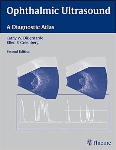
By Cathy W. DiBernardo, Ellen F. Greenberg
ISBN-10: 1588905039
ISBN-13: 9781588905031
This booklet provides the most recent details on utilizing echography to diagnose
lesions and ailments of the attention and orbit. This version is absolutely up-to-date, with a
new bankruptcy on orbital disorder and assurance of the 20-MHz explore for posterior
segment imaging. It presents a accomplished evaluation of the fundamental screening
procedures, descriptions of the symptoms for ultrasound, and assistance on how
to succeed in a correct prognosis of either universal and infrequent medical difficulties in all
areas of the eye.
Features:
- Techniques for diagnosing ailments of the retina,
choroid, vitreous, anterior section, optic nerve, extraocular muscular tissues, and
extra - More than 550 high quality photographs, together with an
multiplied number of anterior phase pictures, that relief the comprehension of
pathology and affliction strategies - Three-dimensional schematics demonstrating sound beam
and probe positions at the eye - Extensive lists of references for pursuing themes in
depth
Ophthalmic Ultrasound: A Diagnostic Atlas will
provide a useful reference for ophthalmologists, ultrasonographers, and radiologists.
Read Online or Download Ophthalmic Ultrasound: A Diagnostic Atlas PDF
Best radiology books

Get The Pathophysiologic Basis of Nuclear Medicine PDF
The second one variation of this publication has been considerably improved to fulfill the calls for of the expanding new pattern of molecular imaging. A separate bankruptcy at the foundation of FDG uptake has been extra. New to this variation are the extra clinically orientated info on scintigraphic experiences, their strengths and barriers when it comes to different modalities.
Comprises every little thing a veterinarian must learn about radiological differential diagnoses. moveable instruction manual layout makes it effortless for daily use Line drawings illustrate radiographic abnormalities in the course of the e-book. special index and vast cross-referencing for speedy and simple use.

Download e-book for iPad: Magnetic Resonance in Chemistry and Medicine by Ray Freeman
High-resolution nuclear magnetic resonance (NMR) spectroscopy and the magnetic resonance imaging (MRI) scanner appear to be worlds aside, however the underlying actual ideas are a similar, and it is sensible to regard them jointly. Chemists and clinicians who use magnetic resonance have a lot to benefit approximately every one other's specialties in the event that they are to make the simplest use of magnetic resonance expertise.
Kevin J. Donohoe, Annick D. Van den Abbeele's Case-based Nuclear Medicine PDF
Compliment for the 1st edition:"Recommend[ed]. .. for beginners and masters alike. it's going to enhance the reader's breadth of data and talent to make sound medical judgements. " - medical Nuclear MedicineIdeal for self-assessment, the second one version of Case-Based Nuclear medication has been absolutely up to date to mirror the newest nuclear imaging expertise, together with state of the art cardiac imaging structures and the newest on PET/CT.
- Radiology of The Sella Turcica
- Radiology of Thalassemia
- Imaging in Biological Research Part B
- Neurosonology and Neuroimaging of Stroke
- Pediatric Ultrasound: Requisites and Applications
Extra info for Ophthalmic Ultrasound: A Diagnostic Atlas
Sample text
20-, 50-, and 100-MHz scleral shells. These shells are less cylindrical to accommodate the probe tip and the movement of the crystal. C I CP Figure 2–29 The ultrasound biomicro- Figure 2–30 The ultrasound biomicroscope (UBM), normal scope (UBM), probe placement. Examination with the UBM. Patient properly positioned with the tip of the probe placed within the fluid-filled scleral shell. angle. The UBM longitudinal scan of the normal angle. Arrow, scleral spur; C, cornea; CP, ciliary processes; I, iris.
Pgs 9/20/06 3:02 PM Page 17 2 ANTERIOR SEGMENT EVALUATION 17 I L F C C T I A Figure 2–18 Cyclitic membrane. Axial immersion image, aphakic eye. Arrow, cyclitic membrane; C, cornea; I, iris. T F S B S P A C B Figure 2–19 Ciliary body mass. (A) Axial scan. Arrow, ciliary body mass; C, cornea; F, fluid, L, lens; P, probe. (B) Transverse scan directly over the lesion. Arrow; ciliary body mass; S, sclera. Figure 2–20 Ciliary body mass. This patient was an HIV-positive 20-year-old African-American male who presented with a large amelanotic ciliary body mass.
Arrow, scleral spur; C, cornea; CP, ciliary processes; I, iris. S AC I L Figure 2–31 The normal ciliary body. Cross-section scan using an ultrasound biomicroscope (UBM). I Figure 2–32 Normal cornea. Ultrasound biomicroscope (UBM) scan through the central axis. The two high reflective lines at the top of the scan correspond to the corneal layers. The corneal stroma (S) is located between the lines. Top arrow, epithelium and Bowman’s membrane; bottom arrow, Descemet’s membrane and endothelium; AC, anterior chamber; I, iris; L, lens.
Ophthalmic Ultrasound: A Diagnostic Atlas by Cathy W. DiBernardo, Ellen F. Greenberg
by George
4.1