By Ruth Dennis, Robert M. Kirberger, Robert H. Wrigley, Frances Barr
This can be the veterinary model of Aids to Radiological Differential prognosis via Chapman and Nakielny. the most goal of the ebook is to supply lists of differential diagnoses for radiological indicators to assist practitioners with logical interpretation of radiographs. as the point of radiological wisdom and services of veterinary basic practitioners is comparatively low (compared with professional radiologists), every one bankruptcy starts with an outline of ordinary radiographic visual appeal. on the way to aid practitioners plan the right way to slim down a listing of differentials or be certain a analysis, feedback for distinction reports and extra diagnostic assessments are indexed. there's a smaller bankruptcy on ultrasonographic indicators that is dependent within the related manner because the radiology part. one other bankruptcy supplies lists of the radiological and ultrasonographic gains of a range of universal stipulations.
Read Online or Download Handbook of Small Animal Radiological Differential Diagnosis, 1e PDF
Similar radiology books
New PDF release: The Pathophysiologic Basis of Nuclear Medicine
The second one variation of this publication has been considerably extended to fulfill the calls for of the expanding new pattern of molecular imaging. A separate bankruptcy at the foundation of FDG uptake has been additional. New to this variation are the extra clinically orientated information on scintigraphic reports, their strengths and boundaries when it comes to different modalities.
Comprises every little thing a veterinarian must find out about radiological differential diagnoses. transportable guide layout makes it effortless for daily use Line drawings illustrate radiographic abnormalities during the e-book. specified index and large cross-referencing for fast and straightforward use.
Get Magnetic Resonance in Chemistry and Medicine PDF
High-resolution nuclear magnetic resonance (NMR) spectroscopy and the magnetic resonance imaging (MRI) scanner appear to be worlds aside, however the underlying actual ideas are an analogous, and it is sensible to regard them jointly. Chemists and clinicians who use magnetic resonance have a lot to benefit approximately every one other's specialties in the event that they are to make the easiest use of magnetic resonance expertise.
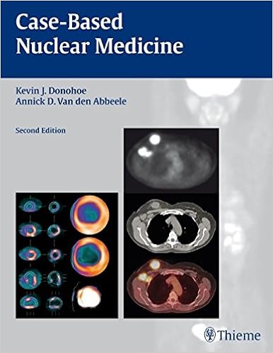
Get Case-based Nuclear Medicine PDF
Compliment for the 1st edition:"Recommend[ed]. .. for beginners and masters alike. it is going to enhance the reader's breadth of information and skill to make sound medical judgements. " - scientific Nuclear MedicineIdeal for self-assessment, the second one version of Case-Based Nuclear drugs has been absolutely up to date to mirror the most recent nuclear imaging know-how, together with state of the art cardiac imaging structures and the most recent on PET/CT.
- MR Cholangiopancreatography: Atlas with Cross-Sectional Imaging Correlation
- Atlas of Perioperative 3D Transesophageal Echocardiography: Cases and Videos
- Radiology Today
- Handbook of Imaging the Alzheimer Brain
- Ultraschallfibel Gynäkologie und Geburtshilfe
Additional resources for Handbook of Small Animal Radiological Differential Diagnosis, 1e
Sample text
7B). The y-direction would then be vertical, in the anterior-to-posterior direction relative to the patient. 7C). In slice selection, the thickness of the excited slice depends on two factors: the strength of the applied magnetic gradient and the bandwidth of the RF pulse sent into the patient. 8). In 2D imaging, slices typically have a thickness between 1 and 10 mm. In 3D imaging, instead of exciting a narrow slice of tissue, a thicker slab of tissue, typically 15 to 40 cm is excited, which uses a wider bandwidth RF pulse or a weaker magnetic gradient applied as the RF pulse is sent into the patient.
The receiver coil’s signal is proportional to the strength of signal coming collectively from the hydrogen nuclei precessing in the tissue sample. In contrast, MRI measures the signal from excited tissues but goes on to localize the MR signal from each voxel in a tissue sample. Chapter 3 will take us from NMR to MRI. There we discuss how signal is measured in each voxel in the human body. Chapter Take-home Points ● Atomic nuclei with either an odd number of protons or an odd number of neutrons have nuclear magnetic dipole moments.
7B). T2 is shorter in fibroglandular tissues than in pure water, CSF, or cystic fluid because the higher concentration of macromolecules in normal breast tissue slows the motion of water molecules, causing them to experience greater magnetic field non-uniformities and therefore dephase more rapidly. 7C). Therefore, their T1 and T2 values are slightly longer than those of fibroglandular tissues but not nearly as long as CSF or cystic fluids. 7D) tend to have longer T1 values because the exchange of energy between hydrogen nuclei and macromolecules is limited, but shorter T2 values than in A-C because of the large magnetic field inhomogeneities maintained by the lattice structure.
Handbook of Small Animal Radiological Differential Diagnosis, 1e by Ruth Dennis, Robert M. Kirberger, Robert H. Wrigley, Frances Barr
by Kevin
4.3


