By Edward I. Bluth
ISBN-10: 1588906108
ISBN-13: 9781588906106
ISBN-10: 1604064684
ISBN-13: 9781604064681
ISBN-10: 3131291427
ISBN-13: 9783131291424
Ultrasonography in Vascular illnesses: a pragmatic method of scientific difficulties is a concise advisor to the newest medical functions of ultrasound in diagnosing ...
summary: Ultrasonography in Vascular illnesses: a realistic method of scientific difficulties is a concise advisor to the most recent scientific purposes of ultrasound in diagnosing
Read or Download Ultrasonography in vascular diseases: a practical approach to clinical problems PDF
Similar radiology books
The Pathophysiologic Basis of Nuclear Medicine - download pdf or read online
The second one version of this booklet has been considerably elevated to satisfy the calls for of the expanding new pattern of molecular imaging. A separate bankruptcy at the foundation of FDG uptake has been extra. New to this variation are the extra clinically orientated info on scintigraphic experiences, their strengths and barriers when it comes to different modalities.
Contains every little thing a veterinarian must learn about radiological differential diagnoses. moveable guide layout makes it effortless for daily use Line drawings illustrate radiographic abnormalities in the course of the e-book. particular index and huge cross-referencing for speedy and simple use.
Ray Freeman's Magnetic Resonance in Chemistry and Medicine PDF
High-resolution nuclear magnetic resonance (NMR) spectroscopy and the magnetic resonance imaging (MRI) scanner appear to be worlds aside, however the underlying actual ideas are an analogous, and it is sensible to regard them jointly. Chemists and clinicians who use magnetic resonance have a lot to profit approximately every one other's specialties in the event that they are to make the simplest use of magnetic resonance know-how.
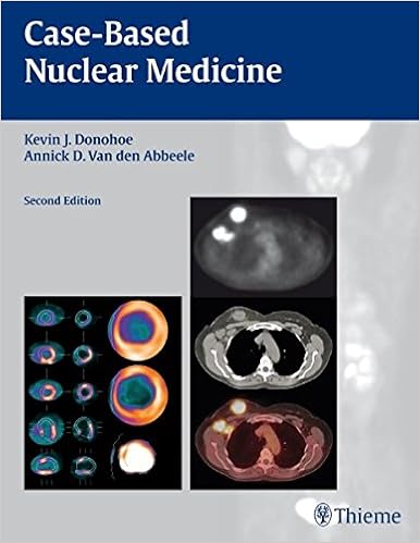
Case-based Nuclear Medicine by Kevin J. Donohoe, Annick D. Van den Abbeele PDF
Compliment for the 1st edition:"Recommend[ed]. .. for newcomers and masters alike. it's going to enhance the reader's breadth of data and talent to make sound medical judgements. " - medical Nuclear MedicineIdeal for self-assessment, the second one variation of Case-Based Nuclear medication has been totally up to date to mirror the newest nuclear imaging know-how, together with state of the art cardiac imaging structures and the most recent on PET/CT.
- Population Monitoring and Radionuclide Decorporation Following a Radiological Or Nuclear Incident
- Biliary Tract Radiology
- Functional Brain Imaging
- Radiology of Influenza: A Practical Approach
Extra info for Ultrasonography in vascular diseases: a practical approach to clinical problems
Sample text
Occasionally, this branch will be visualized in the upper thigh and should not be confused with a PSA neck, a patent needle track, or the arterial limb of an AVF. This vessel does have a relatively low-resistance waveform somewhat atypical of the Figure 3–17 A transverse scan of the right groin in a postcatheterization patient demonstrates partial thrombosis (arrows) of the right common femoral vein (V). qxd 40 8/16/07 2:13 PM Page 40 Ultrasonography in Vascular Diseases A B Figure 3–18 (A) Hyperemic lymph nodes simulating a pseudoaneurysm (arrows).
Note the flow void between the stent and the lumen due to fibrointimal hyperplasia. A stenosis results that is moderately severe by visual assessment. (B) The prestenotic Doppler waveforms are monophasic due to diffuse arterial disease with stenosis proximal to the stent. Peak systolic velocity (PckV) is 88 cm/s. (C) Within the stent stenosis, PckV is slightly more than doubled, at 184 cm/s, indicating hemodynamic significance, but not critical narrowing. (D) Doppler waveforms distal to the stenosis are slightly more damped and lower in velocity (PckV 57 cm/s) than those recorded proximal to the stenosis, suggesting that this stenosis is affecting blood flow.
E) The common femoral vein above the level of the AVF demonstrates normal respiratory phase change (arrow) without evidence of high-velocity pulsatile flow. qxd 8/16/07 2:13 PM Page 39 3 39 Pulsatile Groin Mass in the Postcatheterization Patient A B Figure 3–16 (A) Transverse color Doppler ultrasound of the left groin in a patient with a new bruit status postcatheterization demonstrates a hypoechoic mass in the common femoral artery (A), which obliterates roughly two thirds of the vascular lumen.
Ultrasonography in vascular diseases: a practical approach to clinical problems by Edward I. Bluth
by Daniel
4.4


