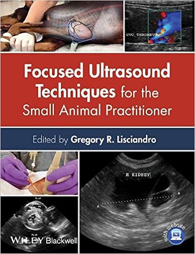
By Gregory R. Lisciandro
ISBN-10: 1118369599
ISBN-13: 9781118369593
ISBN-10: 1118760778
ISBN-13: 9781118760772
Focused Ultrasound ideas for the Small Animal Practitioner offers a hugely sensible advisor to incorporating abbreviated ultrasound tests into the veterinary perform. concentrated point-of-care checks are a great way to speedy observe stipulations and issues no longer with no trouble obvious during the actual examination, laboratory diagnostics, or radiographic findings. Encompassing all of the info had to start acting those concepts, Focused Ultrasound ideas for the Small Animal Practitioner is an invaluable instrument for bettering sufferer results in medical practice.
Covering concentrated assessments in all physique platforms, the publication additionally outlines the rules of interventional radiology, clinical documentation, and the elemental basics of utilizing an ultrasound machine. A significant other site bargains 87 movies of AFAST, TFAST, and Vet Blue examinations with common, irregular, and incidental findings. Focused Ultrasound strategies for the Small Animal Practitioner is a vital buy for veterinary practitioners and experts eager to enforce those recommendations of their veterinary practice.
Content:
Chapter 1 Focused—Basic Ultrasound rules and Artifacts (pages 1–16): Robert M. Fulton
Chapter 2 The stomach FAST3 (AFAST3) examination (pages 17–43): Gregory R. Lisciandro
Chapter three centred or COAST3—Liver and Gallbladder (pages 44–64): Stephanie Lisciandro
Chapter four targeted or COAST3—Spleen (pages 65–79): Stephanie Lisciandro
Chapter five concentrated or COAST3—Kidneys (pages 80–98): Stephanie Lisciandro
Chapter 6 targeted or COAST3—Urinary Bladder (pages 99–109): Stephanie Lisciandro
Chapter 7 targeted or COAST3—Gastrointestinal and Pancreas (pages 110–125): Søren Boysen and Jennifer Gambino
Chapter eight centred or COAST3—Reproductive (pages 126–139): Robert M. Fulton
Chapter nine The Thoracic FAST3 (TFAST3) examination (pages 140–165): Gregory R. Lisciandro
Chapter 10 The Vet Blue Lung experiment (pages 166–188): Gregory R. Lisciandro
Chapter eleven concentrated or COAST3—ECHO (Heart) (pages 189–205): Teresa DeFrancesco
Chapter 12 concentrated or COAST3—Central Venous and Arterial Line Placement, significant Arteries, and Veins (pages 206–221): Scott Chamberlin
Chapter thirteen centred or COAST3—Pediatrics (pages 222–242): Autumn P. Davidson and Tomas W. Baker
Chapter 14 concentrated or COAST3—Eye (pages 243–260): Jane Cho
Chapter 15 targeted or COAST3—Musculoskeletal (pages 261–268): Gregory R. Lisciandro
Chapter sixteen concentrated or COAST3—Cardiopulmonary Resuscitation (CPR), international quickly (GFAST3), and the FAST‐ABCDE examination (pages 269–285): Gregory R. Lisciandro and Andrea Armenise
Chapter 17 Interventional Ultrasound‐Guided approaches (pages 286–303): Søren Boysen
Read Online or Download Focused Ultrasound Techniques for the Small Animal Practitioner PDF
Best radiology books
New PDF release: The Pathophysiologic Basis of Nuclear Medicine
The second one variation of this ebook has been considerably improved to fulfill the calls for of the expanding new development of molecular imaging. A separate bankruptcy at the foundation of FDG uptake has been extra. New to this variation are the extra clinically orientated information on scintigraphic experiences, their strengths and boundaries with regards to different modalities.
Contains every little thing a veterinarian must find out about radiological differential diagnoses. transportable instruction manual layout makes it effortless for daily use Line drawings illustrate radiographic abnormalities in the course of the e-book. exact index and broad cross-referencing for fast and straightforward use.
Download PDF by Ray Freeman: Magnetic Resonance in Chemistry and Medicine
High-resolution nuclear magnetic resonance (NMR) spectroscopy and the magnetic resonance imaging (MRI) scanner appear to be worlds aside, however the underlying actual rules are an analogous, and it is sensible to regard them jointly. Chemists and clinicians who use magnetic resonance have a lot to profit approximately every one other's specialties in the event that they are to make the easiest use of magnetic resonance know-how.
Kevin J. Donohoe, Annick D. Van den Abbeele's Case-based Nuclear Medicine PDF
Compliment for the 1st edition:"Recommend[ed]. .. for beginners and masters alike. it is going to increase the reader's breadth of data and skill to make sound scientific judgements. " - medical Nuclear MedicineIdeal for self-assessment, the second one variation of Case-Based Nuclear drugs has been absolutely up to date to mirror the most recent nuclear imaging expertise, together with state-of-the-art cardiac imaging platforms and the most recent on PET/CT.
- Managing Common Interventional Radiology Complications: A Case Based Approach
- Callen’s Ultrasonography in Obstetrics and Gynecology
- Radiology of the Pancreas
- Biliary Tract Radiology
- Neurosonology and Neuroimaging of Stroke
- Chest X Ray In Clinical Practice
Extra info for Focused Ultrasound Techniques for the Small Animal Practitioner
Example text
The gallbladder wall and its shape should be noted, and the gain may be adjusted based on the echogenicity of its luminal contents for the remainder of the AFAST3 exam. 21). 2C). In low-scoring dogs, one of the most common positive sites is the DH view (along with the CC view). 2C and D). The sonographer should now use the DH view advantageously (less lung [air] interference) as an Keep 25%–33% of the far field as a window into the thorax as you fan through the DH view. 2E). acoustic window (via the liver and gallbladder) into the thorax.
In the event an AFS 1, 2 dog without pre-existing anemia becomes anemic (packed cell volume (PCV) less than 30%), the attending clinician should explore other sites (retroperitoneal, pericardial, pleural, lungs, fracture sites) readily accessible by AFAST3, TFAST3, and the Vet BLUE lung scan and focused musculoskeletal exams at fracture sites for hemorrhage (Chapter 15). AFS 3 and 4 dogs, or big bleeders, will reliably become anemic, predictably 20%–25% below their admission baseline PCV if they had no pre-existing anemia.
2). 2 5 T he A bdominal FA S T 3 ( A FA S T 3 ) E xam Once the acquisition of the gallbladder kissing the diaphragm view is mastered, the sonographer should add on the right-sided cardiac volume status evaluation by generally directing the probe slightly downward (in some patients slightly upward) toward the table top (right lateral recumbency). This builds skills in evaluating for caudal vena caval diameter (as it passes through the diaphragm) and associated hepatic venous distention, using them as markers for right-sided heart status and patient volume status including use in pre- and post resuscitation.
Focused Ultrasound Techniques for the Small Animal Practitioner by Gregory R. Lisciandro
by Brian
4.3