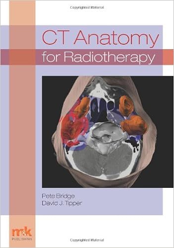
By Pete Bridge
ISBN-10: 1905539541
ISBN-13: 9781905539543
Wisdom of CT anatomy is more and more very important in day-by-day radiotherapy perform, specifically with extra frequent use of cross-sectional image-guided radiotherapy (IGRT) ideas. present CT anatomy texts are predominantly written for the diagnostic practitioner and don't regularly deal with the radiotherapy concerns whereas emphasising constructions that aren't universal to radiotherapy perform. “CT Anatomy for Radiotherapy” is a brand new radiotherapy-specific textual content that's meant to organize the reader for CT interpretation for either IGRT and therapy making plans. it really is appropriate for undergraduate scholars, certified treatment radiographers, dosimetrists and should be of curiosity to oncologists and registrars engaged in remedy making plans. All crucial buildings suitable to radiotherapy are defined and depicted on 3D photos generated from radiotherapy making plans platforms. System-based labelled CT photographs taken in appropriate imaging planes and sufferer positions increase figuring out of relational anatomy and CT interpretation. pictures are observed by way of accomplished observation to assist with interpretation. This simplified method is used to empower the reader to speedily achieve photo interpretation abilities. The ebook can pay certain consciousness to lymph node identity in addition to that includes a distinct part on Head and Neck Deep areas to assist figuring out of universal pathways of tumour unfold. absolutely labelled CT photographs utilizing radiotherapy-specific perspectives and positioning are complemented the place suitable through MR and fusion pictures. a short creation to picture interpretation utilizing IGRT units is additionally lined. the focal point of the ebook is on radiotherapy and a few pictures of universal tumour pathologies are utilised to demonstrate a few suitable irregular anatomy. brief self-test questions support to maintain the reader engaged all through.
Read or Download CT Anatomy for Radiotherapy PDF
Best radiology books
Abdelhamid H. Elgazzar's The Pathophysiologic Basis of Nuclear Medicine PDF
The second one variation of this booklet has been considerably extended to satisfy the calls for of the expanding new development of molecular imaging. A separate bankruptcy at the foundation of FDG uptake has been further. New to this version are the extra clinically orientated info on scintigraphic stories, their strengths and barriers in terms of different modalities.
Comprises every thing a veterinarian must learn about radiological differential diagnoses. moveable guide structure makes it effortless for daily use Line drawings illustrate radiographic abnormalities during the e-book. particular index and large cross-referencing for fast and simple use.
Ray Freeman's Magnetic Resonance in Chemistry and Medicine PDF
High-resolution nuclear magnetic resonance (NMR) spectroscopy and the magnetic resonance imaging (MRI) scanner appear to be worlds aside, however the underlying actual ideas are a similar, and it is smart to regard them jointly. Chemists and clinicians who use magnetic resonance have a lot to profit approximately each one other's specialties in the event that they are to make the easiest use of magnetic resonance expertise.
Read e-book online Case-based Nuclear Medicine PDF
Compliment for the 1st edition:"Recommend[ed]. .. for newcomers and masters alike. it's going to increase the reader's breadth of information and talent to make sound scientific judgements. " - medical Nuclear MedicineIdeal for self-assessment, the second one version of Case-Based Nuclear drugs has been absolutely up to date to mirror the newest nuclear imaging expertise, together with state-of-the-art cardiac imaging structures and the newest on PET/CT.
- Clinical Breast Imaging: A Patient Focused Teaching File
- Atlas of Small Animal CT and MRI
- Procedures in Gastrointestinal Radiology
- Workbook and Licensure Exam Prep for Radiography Essentials for Limited Practice (4th Edition)
- A Concise Guide to Nuclear Medicine
- Oxford textbook of neuroimaging
Additional info for CT Anatomy for Radiotherapy
Sample text
The spermatic cord is a little easier to localise, since it is larger and contains the vas deferens, testicular artery, vein, lymphatic ducts, nerves and surrounding fatty tissues. 4, the vasa deferentia leave the spermatic cords and enter the pelvic cavity. From here, they cross over the external iliac vessels and between the supero-lateral borders of the bladder and the lateral pelvic wall. 3 (30). 4 medial to the seminal vesicles (31). At this point, the vasa deferentia are easier to demarcate due to ampullary dilatation.
1), passing inferiorly and then medially to enter the jejunum. 1 it does so, it passes under the liver. 2). It is the largest gland in the human body and is comprised of two main lobes, the small left lobe and the larger right lobe. The right lobe contains other much smaller lobes known as the caudate and quadrate lobe. The liver is not a major source of primary tumours but due to its rich blood supply it is a common site for metastatic deposits to grow. Sitting under the liver is the pear-shaped gall bladder which stores bile to assist with fat emulsification.
Each kidney is accompanied by an adrenal gland which is located superiorly and medially. 1. Recall that the right kidney is the lower of the two due to the volume of liver above it. Urine is produced in the kidneys by filtering out waste products from the bloodstream. The urine collects in the renal pelvis at the centre of the kidney. From here, the urine passes into the ureters. These are long and take an often tortuous route through the abdomen. It can be difficult to pick out the ureters on individual slices, especially with non-contrast images where they can mimic blood vessels.
CT Anatomy for Radiotherapy by Pete Bridge
by Steven
4.4